
What Is a Brain Matrix? A Complete Guide for Accurate Brain Sectioning
Learn what a brain matrix is, how it works, why it is essential in neuroscience research, and how to choose the right model for reproducible rodent brain slicing in laboratory workflows.
Precision Cutting
Brain matrices guide razor blades through fixed channels to create uniform sections with dependable spacing.
Research Consistency
Consistent slicing supports histology, biochemical analysis, neuroanatomy studies, and pharmacological research.
Custom Solutions
Rapidaccu offers standard mouse and rat models as well as custom cavity sizes and slice intervals.
Definition
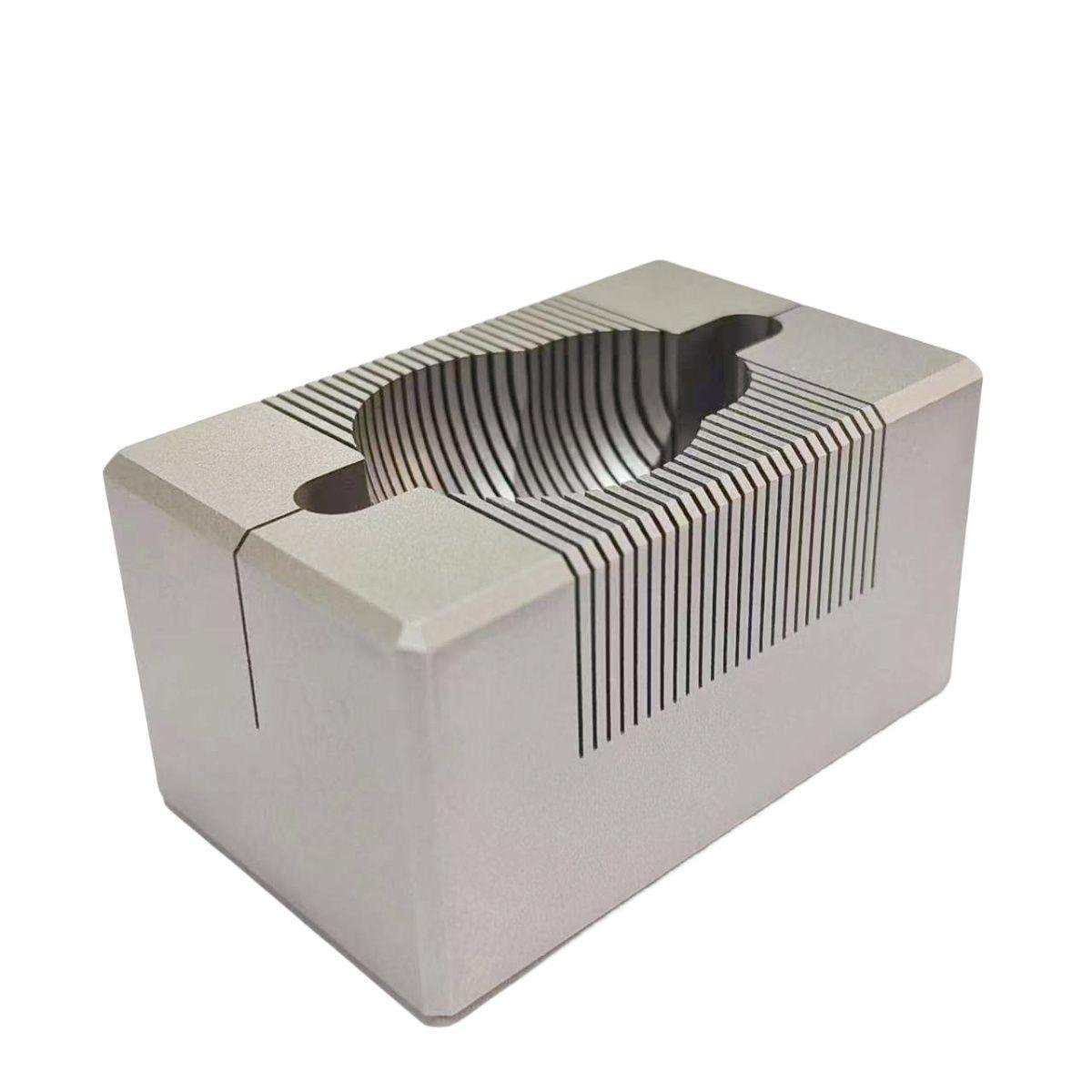
What Is a Brain Matrix?
A brain matrix is a precision laboratory tool designed to hold a brain specimen in position and guide razor blades through fixed channels to produce uniform slices. It is commonly used in rodent research, especially for mouse and rat brains, where reproducibility and anatomical accuracy are critical.
By using predefined blade slots, researchers can create repeatable sections in the coronal or sagittal plane. This reduces variation between samples and helps standardize tissue preparation for downstream analysis.
In practical laboratory workflows, a brain matrix serves as an efficient solution for preparing brain tissue for histology, molecular biology, biochemical assays, and pharmacological evaluation.
Process
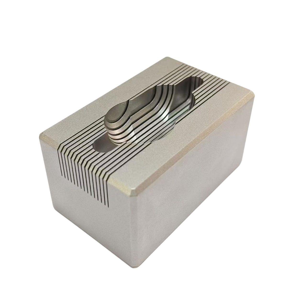
How Does a Brain Matrix Work?
Place the Tissue
The brain specimen is positioned inside the matrix cavity to align with the intended orientation and cutting plane.
Align the Blade
Standard double-edged razor blades are inserted through the matrix channels at fixed intervals.
Create Uniform Slices
The guided channels help produce evenly spaced sections with better repeatability than freehand cutting.
Prepare for Analysis
The resulting tissue blocks can then be processed for staining, imaging, molecular extraction, or regional dissection.
Benefits
Why Is a Brain Matrix Important in Research?
In neuroscience and preclinical studies, small differences in tissue preparation can affect data quality. A brain matrix improves sectioning consistency, helping researchers compare similar anatomical regions across multiple samples.
This level of reproducibility is especially valuable in studies involving region-specific protein extraction, RNA isolation, lesion analysis, or drug distribution assessment.
For laboratories handling routine rodent tissue preparation, a well-made brain matrix can improve workflow efficiency while reducing operator-dependent variation.
Key Advantages
- Improves sectioning accuracy and repeatability
- Supports standardization across experiments and operators
- Helps isolate comparable anatomical regions
- Enhances sample preparation for downstream workflows
- Reduces errors associated with freehand slicing
Use Cases
Common Applications of Brain Matrices
Histological Preparation
Uniform tissue blocks are important before cryostat or microtome sectioning. Brain matrices help prepare samples with consistent dimensions for staining and microscopy.
Biochemical Analysis
Researchers can reproducibly isolate specific regions of the brain for protein, DNA, RNA, neurotransmitter, or metabolite analysis.
Pharmacological Studies
Precise slicing helps evaluate drug distribution, target engagement, and tissue response in defined anatomical coordinates.
Neuroanatomical Research
Consistent brain sections support lesion studies, structural comparison, brain mapping, and region-based investigations in laboratory settings.
Comparison
Stainless Steel vs. Acrylic Brain Matrices
Stainless Steel
- Highly durable for long-term laboratory use
- Autoclavable for sterile workflows
- Corrosion resistant and easy to maintain
- Can be chilled before dissection to help preserve tissue firmness
- Heavier structure provides strong working stability
Acrylic
- Transparent body helps confirm tissue placement visually
- Lightweight and easy to handle
- Suitable for routine laboratory workflows
- Generally not suitable for autoclave sterilization
- Less robust than stainless steel in intensive lab environments
Selection Guide
How to Choose the Right Brain Matrix
Choosing the right brain matrix depends on your species, tissue size, desired cutting plane, slice interval, and sterilization requirements. Mouse and rat studies often need different cavity dimensions, and some protocols call for non-standard spacing for region-specific sampling.
If your lab handles repeated sterilization cycles or intensive use, stainless steel may be the best option. If visual specimen placement is your priority, acrylic may be a practical alternative for selected workflows.
Consider These Factors
- Target species and specimen size
- Coronal or sagittal sectioning orientation
- Required slice thickness or interval spacing
- Cleaning and sterilization method
- Need for standard or customized dimensions
Rapidaccu Advantage
Why Choose Rapidaccu Brain Matrices?
Rapidaccu manufactures high-quality brain matrices for reproducible rodent brain sectioning. Our solutions are designed for laboratory precision, material durability, and workflow flexibility across neuroscience and preclinical applications.
FAQ
Frequently Asked Questions
What is a brain matrix used for?
What species are brain matrices commonly made for?
What is the difference between coronal and sagittal sectioning?
Should I choose stainless steel or acrylic?
Can Rapidaccu provide custom brain matrices?
What kind of blades are used with a brain matrix?
Need a Standard or Custom Brain Matrix?
Talk to Rapidaccu about your rodent brain sectioning requirements and get a solution designed for your research workflow.
Contact Information
Get in Touch
Address
1/F and 2/F, No.4 Building, Fulongte Industrial Park, No.5 Huaxing Road, Dalang Street, Longhua District, Shenzhen City, Guangdong Province, China.
Request Quote